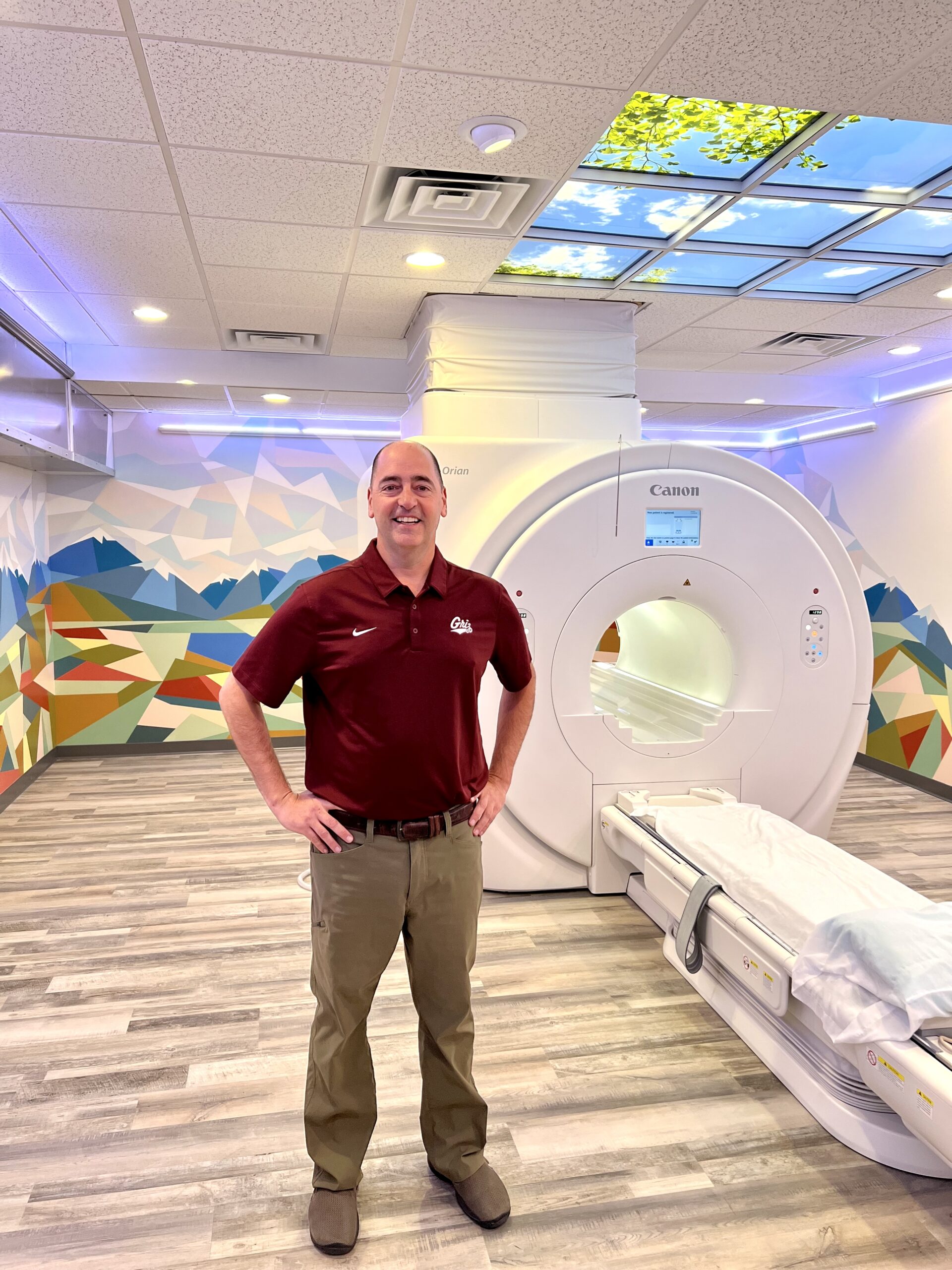
High-quality MRIs
without all the hassle.
Super simple and freaky fast.
Missoula’s fastest
MRI scan experience
MRI (Magnetic Resonance Imaging) works with a large magnet, radio waves, and a computer to produce detailed images of the inside of the body. It uses electromagnetic energy rather than ionizing radiation or any kind of radioactive material. MRI provides physicians with a powerful tool to examine virtually any structure in your body, including bones, muscles, connective tissue, and organs such as the heart, lung, or kidneys.
We will deliver all your images and reports to your provider, whether they’re across town or across the country.
Did you know our MRI machine is just plain better?
Wide Bore “Open Bore” MRI
With an opening that’s nearly 30% larger than the older traditional narrow bore MRIs that can be found in the Missoula area hospitals, our wide-bore MRI (also called Open Bore MRI) provides the ultimate in patient comfort during the examination.
Our wide bore MRI scanner has the widest bore in the region with a 71 centimeter bore opening. That extra 11 centimeters can make all the difference for many patients. Those who are claustrophobic often find that the wider bore is less stressful than the older traditional, narrow bore MRIs that can be found in the Missoula area hospitals.
The Open Bore MRI combines the benefits of an open MRI with increased space inside the bore area of the machine and has even more headroom than the traditional Open MRI machines.
Because our MRI system delivers both a high magnet strength for higher quality imaging, the overall MRI procedure time is shorter in duration than the traditional Open MRI.
Choosing our Open Bore MRI system at Montana Imaging Center, your patient preparation time is reduced, there are faster scan times in the MRI machine, patient comfort is increased, and image quality for your referring doctor is high.

WE HAVE THE FASTEST MACHINE
With the widest opening and fastest speed, our machine is the best in the state.
Need an arthrogram?
We can do those.
An Arthrogram is a diagnostic test which examines the inside of a joint (e.g. shoulder, knee, wrist, ankle) to assess an injury or a symptom you may be experiencing. The test is done by first injecting contrast medium (or “dye” as it is sometimes called) into the joint, which outlines the soft tissue structures in the joint (e.g. ligaments and cartilage). It makes them clearer to see on the images or pictures that will be taken of the joint. This is done via ultrasound or x-ray guidance at The Montana Imaging Center. Ultrasound and X-ray help to guide the placement of the needle containing the contrast medium.
This is then followed by a magnetic resonance imaging (MRI). While an MRI without the use of contrast medium can provide information on the soft tissue structures, using contrast medium with MRI Arthrogram (injecting into the joint) may provide more information about what is wrong with the joint.
What can we scan?
- Brain
- Cervical Spine (Upper/Neck Portion)
- Upper Extremities (Shoulder, Wrist, Hand, Finger, etc.)
- Soft Neck Tissue
- Sacrum/Coccyx
- Orbits
- Thoracic Spine (Chest Portion)
- Lower Extremity (Hip, Knee, Ankle, Foot, etc.)
- Pelvis Soft Tissue
- Neck/Carotid
- Chest
- Lumbar Spine (Lower Portion)
- Abdomen (MRCP, Kidneys, Adrenal Glands, Liver)
- Bony Pelvis
- Breast Implants
Get in,
get scanned,
get back out.
Get your scan today and have the report back in 72 hours.


